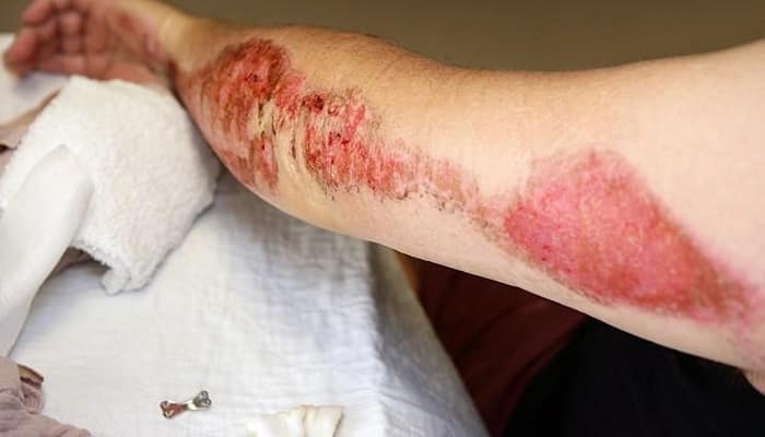
Skin necrosis, called necrosis, is local tissue death that occurs due to tissue damage or infection of skin wounds. This may be the result of a thermal irritant – it can occur after a severe burn or frostbite. This may also be a consequence of improper care of bedsores.
Necrosis is initially manifested by swelling and pain, and later by decreased sensitivity. Physical examination plays an important role in the diagnosis of necrosis. If the wound is infected with bacteria, treatment is with antibiotics or chemotherapy. To limit the development of necrosis, it is necessary to spotless the wound.
What does skin necrosis look like?
The skin turns brownish-black. The area where necrosis occurred is swollen and warm. The patient’s temperature often rises. Mummification occurs, i.e., the formation of a crust and the drying of dead tissue. Hemorrhagic epidermal blisters appear. If left untreated, dry necrosis can progress to wet necrosis, and then ulcers form in the gangrenous tissues, oozing brown discharge with an unpleasant odor. Skin necrosis manifests itself in the initial stage as pain, and with advanced necrosis, a complete loss of sensitivity of dead skin occurs. Rapid progression characterizes the disease.
When does skin necrosis occur?
Skin necrosis occurs in a patient due to tissue damage or wound infection. One of the most common causes of its occurrence is a thermal irritant. Skin necrosis develops after a burn. In a third-degree burn, necrosis affects the dermis with nerves and vessels of the skin, as well as subcutaneous fat. The skin becomes hard and dry, gray-white to brown. Fourth degree burns are characterized by necrosis reaching deeper tissues. Skin necrosis also occurs after injuries caused by low temperatures. With third-degree frostbite, necrosis impacts the superficial layer of the skin, and with fourth-degree frostbite, deep necrosis occurs.
Necrosis can be a consequence of improper care of bedsores in bedridden people and crushing of the skin. Skin necrosis can also occur as a result of an abnormal reaction of the body after taking warfarin. The etiological factors of this condition include: protein C deficiency, hypersensitivity and direct toxic effects of warfarin. Changes appear 3–6 days after starting warfarin treatment.
Skin necrosis can occur after surgery when the wound becomes infected. Then the group at increased risk of necrosis includes: patients with atherosclerosis and other vascular diseases, elderly people, smokers, exhausted people, alcohol abusers, drug addicts, people with reduced immunity and high levels of cholesterol in the blood, with kidney failure, lung disease, liver disease, against the background corticosteroid therapy. People with diabetes are especially susceptible to skin necrosis and may develop diabetic foot. Then, if treatment is not started promptly, it may even lead to amputation of the foot. Diabetics should avoid situations that could cause injury to their feet and be very careful about their hygiene.
Skin necrosis – treatment
Skin necrosis is a disease that requires immediate treatment, as it can contribute to the development of a generalized infection and, as a result, lead to shock, multiple organ failure and even death. Treatment of skin necrosis is aimed at thorough treatment of the wound – excision of the edges of the wound, cleaning, and removal of foreign bodies. Advanced forms require skin transplantation. If the presence of bacteria is detected, antibacterial therapy is necessary. Additionally, painkillers and drugs that improve blood flow in the vessels are used.
A popular method of treating undeveloped necrosis is ultrasound. They spare healthy tissue and destroy bacterial and fungal flora in the wound. Another method is special hydrogel dressings. By replacing the wound environment with a moist one, they promote the resorption of necrosis. In addition, they stimulate cells to produce enzymes that cleanse the wound. They stimulate the mechanisms of autolysis (digestion of dead cells) in the wound, as well as the granulation process.
A fairly popular solution recently is larvotherapy, which is used especially for clearing necrosis in patients with diabetic foot. The treatment uses blowfly larvae, which are grown in laboratories in compliance with the principles of sterility. The larvae not only cleanse the wound of necrotic tissue, but also reduce the odor and number of secretions oozing from the wound.
Patients with skin necrosis should take care of the general good condition of the body. Therefore, they should drink plenty of fluids and eat a high-calorie, protein-rich diet, which allows the damaged tissue to heal faster. Additionally, they need to take vitamins and microelements that increase immunity. It is also important not to put any weight on the affected limb and avoid possible infection.


